The integration of artificial intelligence into healthcare is no longer a future concept; it is a present-day reality, particularly within the field of medical imaging. For students, researchers, and professionals in biomedical engineering and computer science, understanding how to apply machine learning to oncology is becoming a critical skill. A recent workshop led by Professor Habib Zaidi at Óbuda University highlighted the practical pathways for using AI to improve cancer diagnosis and therapy, offering a clear view of where the industry is heading.
This article synthesizes the key insights from that event, focusing on the technical application of AI in hybrid imaging systems and its role in supporting clinical decision-making.
Understanding the Core Technologies: PET, SPECT, and CT
Effective application of AI in medical imaging begins with a firm grasp of the underlying technologies. Professor Zaidi’s workshop emphasized the role of hybrid systems, which combine functional and anatomical data to provide a more complete diagnostic picture. The primary modalities discussed were:
- Positron Emission Tomography (PET): This nuclear medicine technique visualizes metabolic processes at the cellular level. It is highly effective for detecting cancerous activity, as tumors often exhibit elevated metabolic rates.
- Single-Photon Emission Computed Tomography (SPECT): Similar to PET, SPECT provides detailed, layered visualizations of organ function by tracking the distribution of radiotracers.
- Computed Tomography (CT): CT scans provide high-resolution structural images of the body’s internal anatomy.
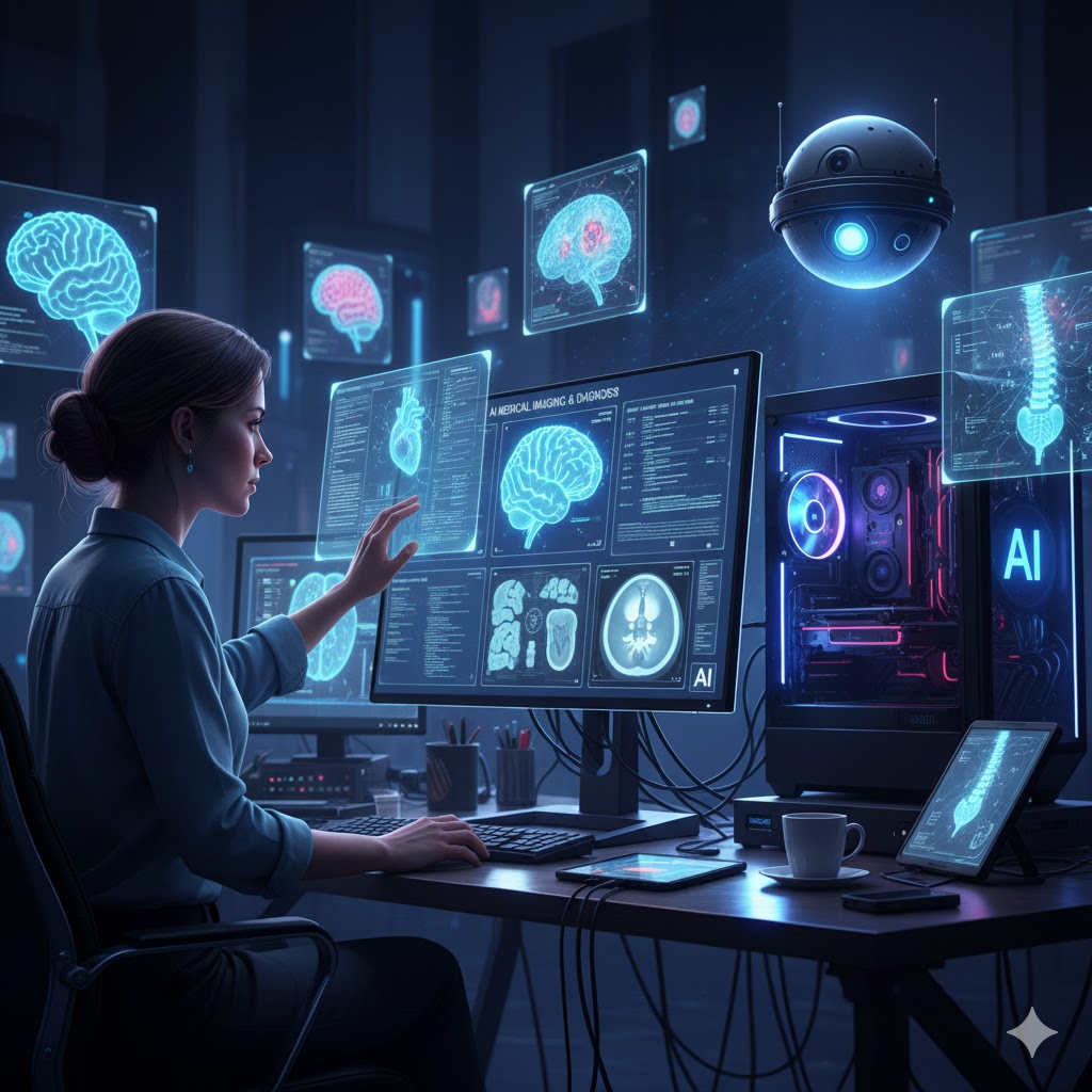
When combined, systems like PET-CT and SPECT-CT allow clinicians to see both the structure of a potential tumor (from CT) and its metabolic activity (from PET/SPECT). This fusion is where AI provides significant value, by analyzing these complex, multi-layered datasets to identify patterns that might be missed by the human eye.
The Role of Artificial Intelligence in Image Analysis
Artificial intelligence, particularly deep learning, is not intended to replace the physician. Instead, it functions as a powerful support tool. The workshop at Óbuda University detailed several key areas where AI algorithms are being applied:
1. Automated Detection and Segmentation
One of the most time-consuming tasks for a radiologist is identifying and outlining suspicious regions across hundreds of image slices. AI models can be trained to automate this process. By learning from vast datasets of annotated images, these algorithms can:
- Flag potential lesions for closer review.
- Segment tumors with high precision, which is essential for accurate volume measurement and treatment planning.
- Reduce the risk of human error or fatigue-related oversight.
2. Image Quality and Artifact Reduction
The quality of a diagnostic image is paramount. Even minor inaccuracies during image reconstruction can lead to artifacts that obscure details or mimic pathology. AI algorithms can enhance image quality by:
- Reducing image noise, which allows for lower radiation doses without sacrificing diagnostic clarity.
- Correcting motion artifacts that can occur during scans of the chest or abdomen.
- Improving the resolution of low-quality scans, making them more useful for diagnosis.
3. Multi-Modal Image Registration and Comparison
Patients often undergo multiple scans over the course of their treatment. Comparing a current PET-CT scan to one from six months prior is a complex task of spatial alignment. AI excels at registering these images, allowing for precise, automated comparison to track disease progression or response to therapy.
Minimizing Risk: Radiation Dose Optimization
A critical concern in medical imaging is the patient’s exposure to ionizing radiation. The workshop highlighted how AI contributes to the principle of ALARA (As Low As Reasonably Achievable). By improving reconstruction algorithms, AI can generate diagnostic-quality images from significantly less raw data. This means that for a CT or PET scan of the brain, heart, or full body, the radiation dose can be minimized while preserving the diagnostic accuracy required for effective cancer diagnosis.
The Future: Personalized Computational Models
Professor Zaidi’s central message points toward a future of personalized medicine. The goal is to move beyond simply identifying a tumor to creating a computational model of an individual patient’s disease. AI is the engine that will power these models, integrating imaging data with genetic information and other biomarkers. This approach will allow clinicians to:
- Predict how a specific cancer will progress.
- Simulate the potential outcomes of different treatment strategies.
- Make optimal, data-driven decisions for each patient.
Advancing Your Career in AI and Medical Imaging
The demand for professionals who can bridge the gap between advanced imaging physics and machine learning is growing rapidly. Institutions like Óbuda University are at the forefront of this field, fostering research and education that addresses these complex challenges. For those looking to contribute to the future of cancer diagnosis and therapy, developing expertise in these areas is a strategic career move.
Engaging with academic research and specialized programs is the most effective way to gain the necessary skills. If you are interested in contributing to this innovative field, consider exploring the research opportunities and academic programs offered by leading institutions.
Explore academic programs in engineering and technology to build a foundation for a career in medical AI.
Conclusion
The application of artificial intelligence in medical imaging is creating new pathways for improving cancer diagnosis and therapy. By enhancing the analysis of PET, SPECT, and CT data, AI supports clinicians in making more accurate and timely decisions. The key takeaway from the work being done at Óbuda University is clear: AI is a collaborative tool that, when combined with expert medical knowledge, has the potential to significantly advance patient care.
For researchers and students, the opportunity to contribute to this transformation is now. Learn more about international research collaborations and take the next step in your professional development.